
Premium Edition
The fluorescence channel
A cooled back-illuminated sCMOS camera captures 2048 × 2048 pixels at up to 40 fps, with sensitivity below 3 nM IR-780 iodide and 70 µm × 125 µm resolution at skin level. Real-time 2D imaging runs at 20 fps in coronal, sagittal or any intermediate view, co-registered with the photoacoustic volume.
Excitation tunes in 1 nm steps across 460–800 nm, the practical equivalent of 340 narrow-band excitation filters. An eight-filter wheel covers emission from 510 to 995 nm, with two slots free.
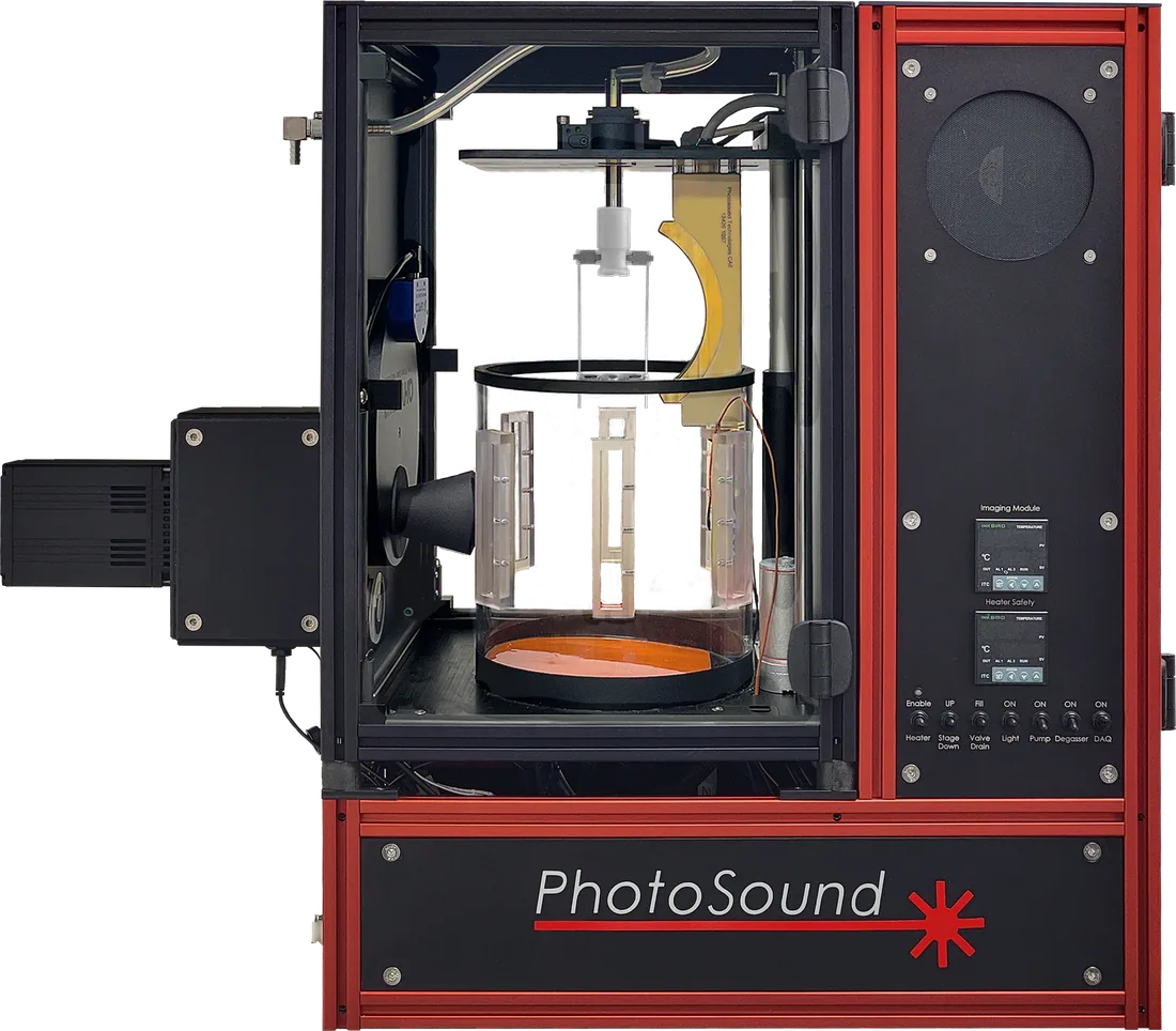
Specifications
| Type | 3D high-resolution deep tissue molecular, physiological and anatomical imaging | Subcutaneous and skin imaging |
|---|---|---|
| Spatial resolution | 150 µm × 150 µm; 150 µm × 420 µm | Transverse planes; sagittal and coronal planes |
| Molecular imaging sensitivity | 100 nM ICG | In vitro tests, CNR = 2 |
| PA excitation range | 460–1320 nm | |
| Detection points per scan | > 69,000 | Single scan, 360° azimuthal rotation |
| Detector configuration | Curve-linear array | Cylindrical focusing |
| Detector central frequency | 6 MHz ± 10% | T/R measurements, optimised sensitivity in receive mode |
| Number of array elements | 96 | Wide-angle 3D imaging transducers |
| PA signal digitizer | LEGION ADC | 12-bit, 256 parallel channels, up to 400 Hz frame rate, 40 MHz sampling, programmable amplifier 46–91 dB |
| Type | Real-time 2D or 3D molecular imaging, co-registered with the PA channel | Real-time 2D in coronal, sagittal or any intermediate view at 20 fps |
|---|---|---|
| Spatial resolution | 70 µm × 125 µm | At skin level of a live test subject |
| Molecular imaging sensitivity | < 3 nM IR-780 iodide | 780 nm excitation, CNR = 4.8 |
| FL excitation range | 460–800 nm | |
| Excitation linewidth | < 1 nm | 1 nm tuning step, equivalent to 340 extremely narrow-band excitation filters |
| Emission filter set | 8 filters, 510–995 nm | Programmatically controlled filter wheel, 2 spare slots |
| Detector type | Back-illuminated sCMOS | High-sensitivity cooled scientific camera |
| Bit depth | 16-bit | |
| Number of pixels | 2048 × 2048 | 19.5 µm pixel resolution |
| Max frame rate | 40 fps | |
| Dynamic range | 86 dB | |
| Quantum efficiency | 95% @ 600 nm | 30–95% across 400–900 nm |
| Readout noise | 1.2 e⁻ | Low readout noise for high frame rate applications |
| Dark current | < 0.008 e⁻/pixel | For 50 ms or shorter exposures |
| Tunable wavelength range | 650–1320 nm and 460–649 nm | |
|---|---|---|
| Pulse repetition frequency | 20 Hz | |
| Pulse energy | > 130 mJ @ 700 nm; > 10 mJ @ 500 nm | Before fiber bundle transmission |
| High-energy excitation @ 1064 nm | > 350 mJ | |
| Energy meter | Real-time automatic pulse energy measurement | |
| Fast wavelength switching | Any wavelength in 650–1320 nm or 460–649 nm every 50 ms | |
| Chassis type | Mobile | On wheels, positioned next to the image acquisition unit |
| Dimensions (L × W × H) | 68 cm × 44 cm × 89 cm | |
| Power requirements | 120, 208 or 240 VAC, single phase 50/60 Hz, < 1.5 kVA |
| Standard scan time | 36 s | 360° azimuthal rotation, 720 data frames |
|---|---|---|
| Scan types | Continuous azimuthal rotation or reverse scans (≤ 360°) | Time-limited to 10 min |
| Excitation sequence | Single wavelength; linear or custom wavelength sweep | Spectral unmixing presets for molecular, physiological and anatomical imaging |
| Max single-scan 3D image | 50 mm × 50 mm × 30 mm | |
| Whole-body imaging | Stack of 3D volumes, manual axial positioning | 10 mm steps, 40 mm positioning range, 50 × 50 × 70 mm total imaging range |
| In vivo imaging subjects | Mice, rats (< 200 g) | Fur should be shaved or depilated from the studied section before imaging |
| Max weight of test subject | 0.5 kg | |
| Coupling medium | DI water | Subject submerged under anesthesia, degassing enabled |
| Environment temperature control | 20–40 ± 0.5 °C | Controlled heating and circulation of the coupling liquid |
| Test subject monitoring | Visual monitoring with a camera | |
| Laser safety | Light-tight imaging chamber, laser interlocks | No eye protection required |
| Chassis type | Benchtop | |
| Dimensions (L × W × H) | 78 cm × 35 cm × 70 cm | 55 cm × 35 cm footprint |
| Power requirements | 208–240 V 4 A or 120 V 8 A, 50/60 Hz |
| Form factor | Desktop mid-tower or Mini ITX case | |
|---|---|---|
| Configuration | High-performance Nvidia GPU, SSD, Windows 10/11 | 1440p or higher monitor, keyboard, mouse |
| Imaging software | TriTom Imaging Suite; 3D Slicer | Acquisition, reconstruction and molecular imaging; visualisation and analysis of 3D volumes |
| Data formats | Scan data: raw, mat. 3D image: vtk, mat. 2D image/video: raw, mat, png, tif, mp4 |
| Transmission | > 70% | |
|---|---|---|
| Excitation spot, axial size | 30 mm | |
| Length | 2 m |
Accessories included
| Gas anesthesia system | Mice and small rats; includes animal induction chamber | |
|---|---|---|
| Mouse restrainer | B-type for the abdominal region and legs; H-type for the thoracic region, head and neck | |
| Microcuvette holder | Scans up to ten 50 µL cuvettes of liquid sample, quick setup | |
| Microcuvettes | Cylindrical PTFE, 0.8 mm ID, 50 µm wall thickness, for 50 µL samples | |
| Coupling liquid containers | Fill and drain the image acquisition unit | |