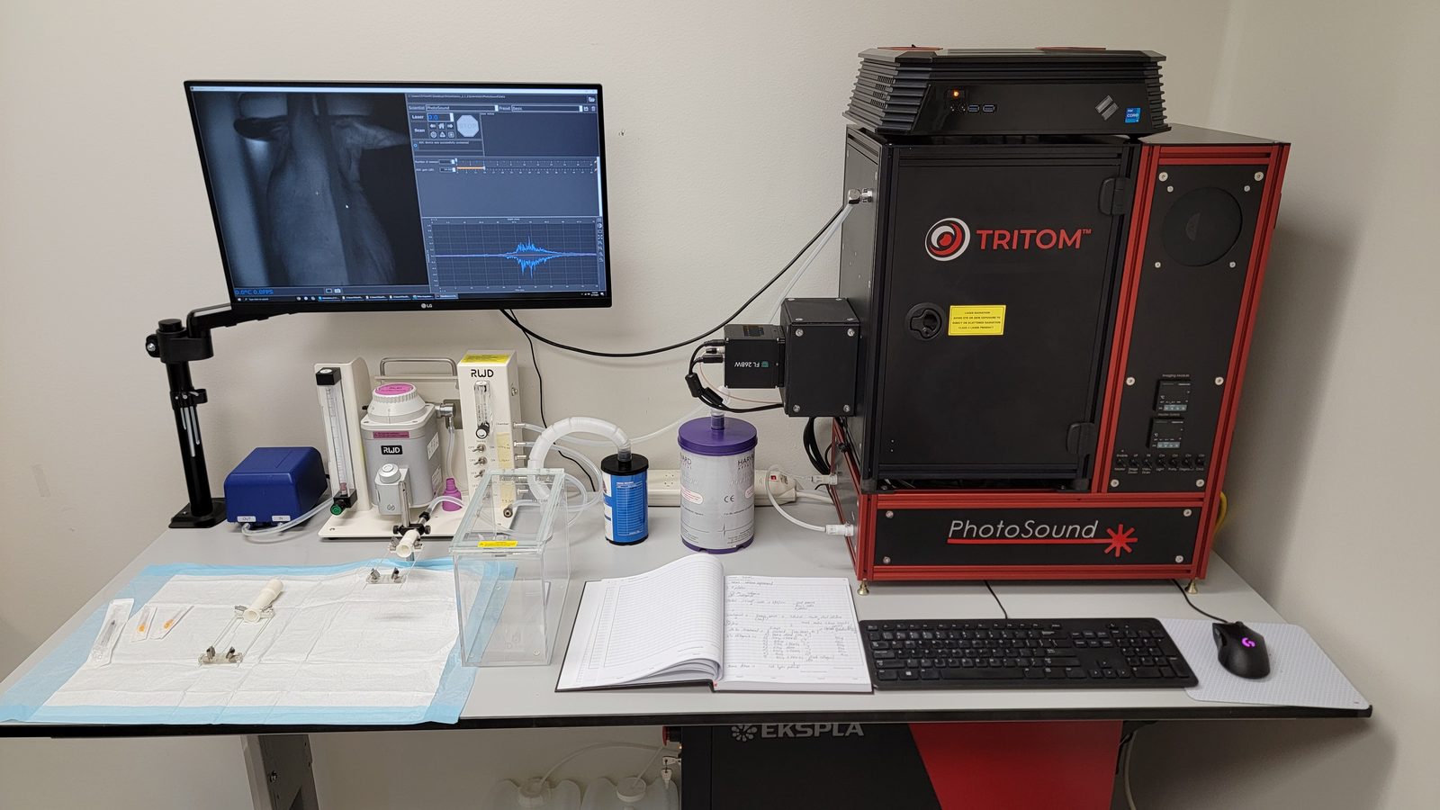
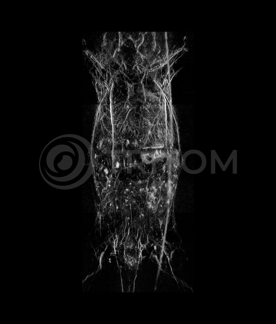
The instrument, an installation, and what comes out of it.
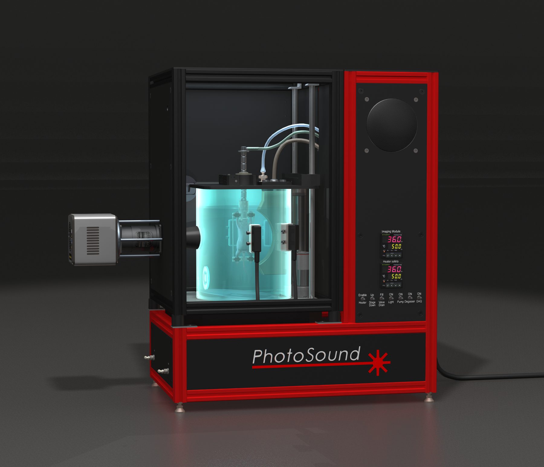
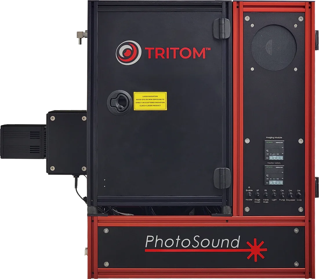
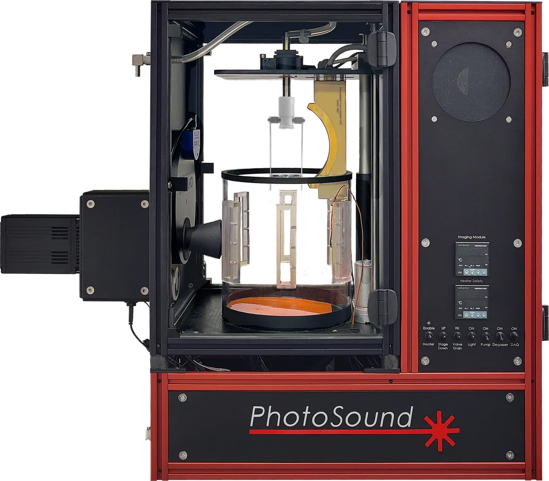
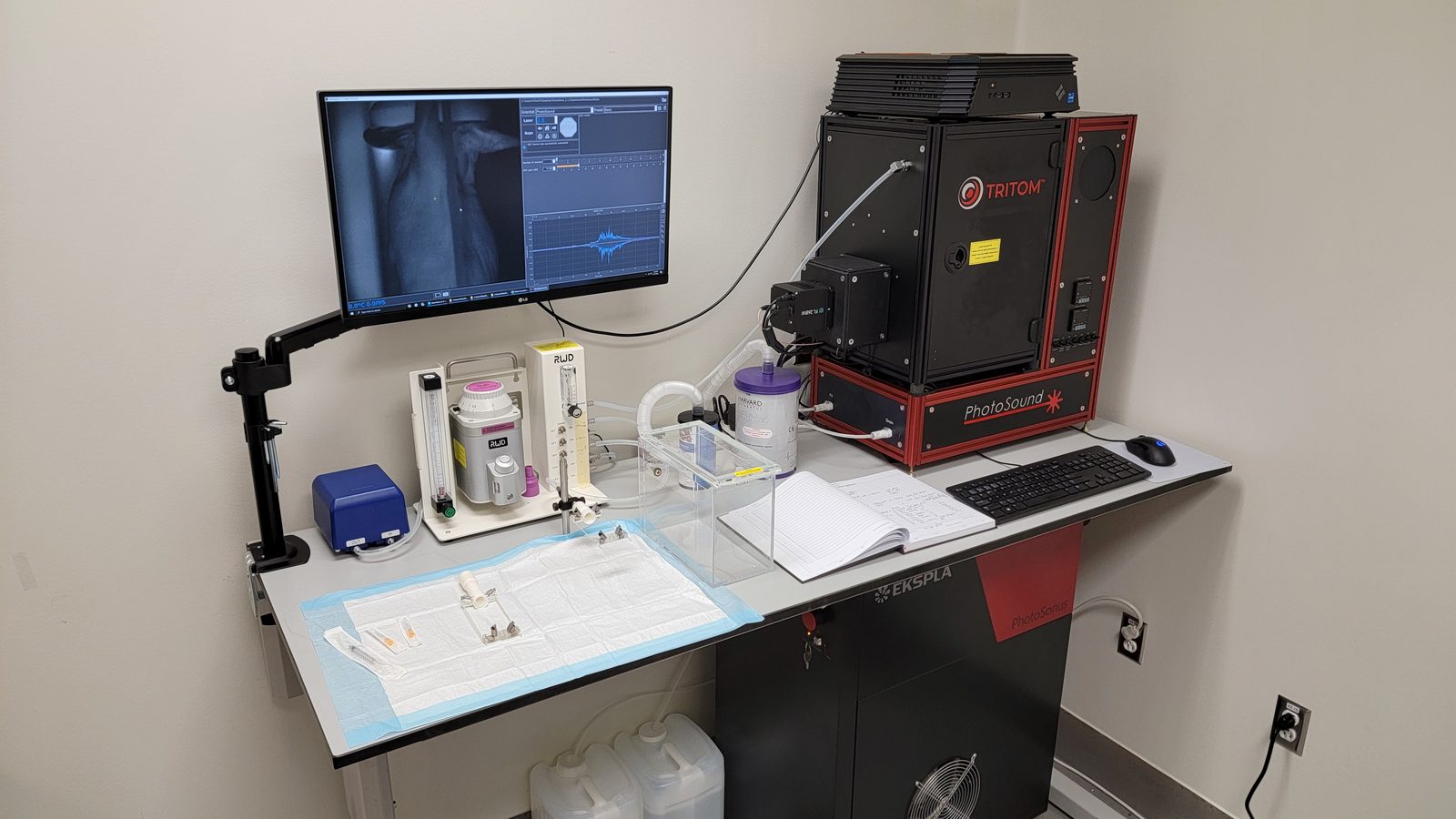
The imaging chamber during a scan, with the subject submerged in temperature-controlled DI water.Chamber closed. Laser interlocks and a light-tight enclosure mean no eye protection is required.Chamber open, with a holder mounted above the coupling liquid vessel.A complete installation: instrument, control station, anesthesia system and animal prep area.The 55 × 35 cm footprint leaves working room on a standard bench.Whole-body maximum intensity projection: circulatory system and internal organs, no contrast agent.